

Facts, figures, and myths: focus on the gut
Research on the gut and its microbiome has literally exploded over the past decade and a half: on the topic of the gut microbiome and disease alone, there are now well over 40,000 scientific publications, with steadily increasing funding and attention. When it comes to the gut, there are also a number of myths that persist despite increased research.
The widely held belief that the intestine has the surface area of a tennis court (often cited as 180 to 300 m²) is one of those myths that was debunked by a new measurement conducted in 2014 using more precise microscopic methods [3]. The actual average mucosal surface area of the digestive tract is only about 32 m², of which approximately 2 m² is accounted for by the large intestine. This puts it closer to half a badminton court than a tennis court. Still, it is impressive for an organ that fits inside our abdomen when folded up.
To remind ourselves why our body needs this seemingly oversized surface—consisting of the mucous membrane (mucosa), connective tissue (submucosa), muscle layer (muscularis), and outer layer (serosa)—here is a brief illustration.
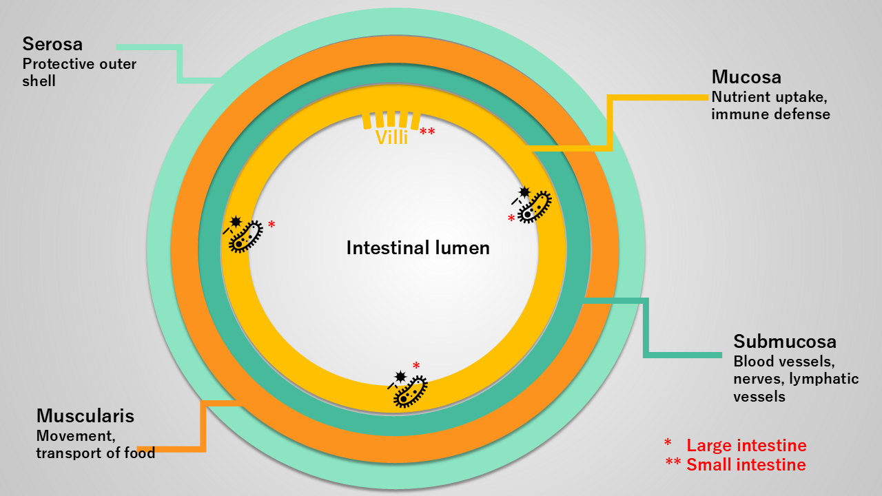
So much for the basic structure for now, but how does a disruption of this wonderfully coordinated system manifest itself, and what role do mast cells play—since they are of particular interest to us? In the case of the following five clinical pictures, it is worth taking a closer look at exactly what can go wrong...
MCAS and the microbiome: a constant dialogue
The mucosa—the innermost layer of the intestinal wall—is not only crucial for nutrient absorption but also for defending against harmful bacteria and toxins. This is where the much-discussed mucosal immune system is located, along with vast numbers of mast cells.
Although mast cells in the gut react directly to microbial signals from their immediate surroundings, communication actually flows in both directions. Messenger substances such as histamine and tryptase, which are released by mast cells, in turn influence which bacteria can colonize the gut [4].
The study by Krausfeldt et al. [2] mentioned at the beginning demonstrates just how close this connection actually is: in patients with systemic mastocytosis, an altered composition of certain bacterial groups (Firmicutes and Bacteroidetes) was found that was directly linked to tryptase levels, a classic marker of mast cell activity. It is, therefore, a constant dialogue that can spiral out of control in either direction if the balance is disrupted.
Important to note: In mast cell activation syndrome (MCAS), unlike in systemic mastocytosis, tryptase levels in the blood are not persistently elevated in many patients; thus, a normal level does not rule out the condition.
And at this point, it is important to distinguish between correlation and causation. If one were to assume that the altered bacterial composition is caused by elevated tryptase levels, one could conclude that individuals with MCAS who have low tryptase levels should not exhibit this altered bacterial composition.
However, the study merely shows that tryptase levels and changes in the microbiome occur together (correlate), which does not necessarily mean that tryptase itself drives the change in the microbiome. It could just as easily be that both are influenced by a third factor, such as overall mast cell activity or the total disease burden. In this context, tryptase is merely one measurable indicator, not the cause. It would therefore be interesting to extend this study design to include individuals with MCAS rather than exclusively those with systemic mastocytosis.
Histamine intolerance and the gut: when bacteria come into play
Many of you may be familiar with this: probiotics are always a bit of a gamble—they can go wrong very quickly and cause or worsen symptoms. To protect yourself from expensive mispurchases, you should look very closely at the list of strains included.
Some bacterial strains, such as Morganella morganii or Enterococcus faecalis, produce histamine themselves, while others, such as Klebsiella pneumoniae, help break it down. Normally, there is a finely balanced equilibrium between the strains, which can easily be disrupted in the event of dysbiosis. A small pilot study of individuals with histamine intolerance accordingly found an altered composition of precisely those bacterial groups that include known histamine producers [5].
However, the actual breakdown of histamine is primarily carried out by our body’s own enzyme, DAO (diamine oxidase). DAO is produced mainly in the intestinal mucosa, which is why a compromised intestinal barrier has a double impact: more histamine from both outside and inside, and at the same time, less capacity to break it down. This makes it all the more important, in cases of histamine intolerance, to specifically assess the state of one’s own gut health.
Autoimmune diseases and the intestinal barrier: a matter of tolerance
The intestinal barrier and our immune tolerance—that is, the immune system’s ability to distinguish between harmless and dangerous substances—are closely linked. Here, too, the two-dimensional nature of the process becomes apparent: certain metabolites produced by our gut bacteria—most notably short-chain fatty acids (SCFAs)—specifically promote those immune cells (regulatory T cells) that curb excessive reactions directed against the body itself [6].
Interestingly, the receptor involved in this process, GPR109A, is the same one we have already encountered in our SCFA article, which focused on its mast cell-suppressing effect. So, the very same binding site regulates several immune mechanisms in the gut at once. If this balance is disrupted, it is now being discussed as a possible factor in several autoimmune diseases. However, exactly how this can be measured and interpreted in individual cases is more complicated than it seems at first glance, so I will address this in more detail in a later article.
POTS: the vagus nerve as a link
When we examine the gut, there is one thing we cannot ignore: the gut-brain axis, connected via the vagus nerve. This important link between the gut and the autonomic nervous system works in both directions. Sensory fibers transmit signals from the gut to the brain, while other fibers travel in the opposite direction to control gut motility.
This is precisely where a possible explanation for postural orthostatic tachycardia syndrome (POTS) comes into play: if autonomic regulation is disrupted, intestinal transit time often changes as well—and, indirectly, which bacteria can colonize the gut. A recent study [7] did indeed find significant differences in microbiome composition between individuals with POTS and healthy individuals; however, an earlier, smaller pilot study reached the opposite conclusion [8]. This is a field of research that is still in its infancy.
Endometriosis and the estrobolome
The collective term for all gut bacteria involved in estrogen metabolism is “estrobolome,” and it sounds so beautifully causal… but here comes a bitter disappointment for anyone expecting a clear, significant link between the gut and endometriosis.
After several smaller studies had suggested that the bacterial composition of the gut might also influence hormonal balance (for example, [9] or [10]), a large-scale cohort study of 1,000 women dispels this hypothesis [11]. No difference in the microbiome was found between women with and without endometriosis.
The authors themselves, however, provide a plausible explanation for this: they had lumped together different subtypes of the disease, ranging from the superficial to the deep-infiltrating form. In keeping with endometriosis’s reputation as the “chameleon” among diseases—a condition that manifests so individually and in so many different ways—it simply defies a blanket answer to the microbiome question. Initial studies that distinguish by subtype are indeed finding differences again. In deep-infiltrating endometriosis, for example, there is a significantly reduced bacterial diversity, while in the ovarian form, there is altered activity of enzymes involved in estrogen metabolism [12]. This is further evidence of how important it is to take a close look at this disease.
An open question to conclude
Five very different conditions, and yet the gut emerges as a possible contributing factor in most of them—even if, as we have just seen with endometriosis, it is not always as clear-cut as it seems at first glance.
The intriguing question that remains is: What do I do with this knowledge now? The next articles in this series might help you answer that question. They will explore whether it makes sense to have your gut examined, and if so, which tests to get and what the results actually mean.
References
[1] Buhner S, Schemann M. Mast cell-nerve axis with a focus on the human gut. Biochim Biophys Acta. 2012 Jan;1822(1):85-92. doi: 10.1016/j.bbadis.2011.06.004. Epub 2011 Jun 17. PMID: 21704703.
[2] Krausfeldt L, Cao V, Rodrigues R et al. Evidence for dysbiosis in the gut microbiome of patients with systemic mastocytosis. Journal of Allergy and Clinical Immunology: Global, 2025; 5. doi: 10.1016/j.jacig.2025.100578
[3] Helander HF, Fändriks L. Surface area of the digestive tract - revisited. Scand J Gastroenterol. 2014 Jun;49(6):681-9. doi: 10.3109/00365521.2014.898326. Epub 2014 Apr 2. PMID: 24694282.
[4] Carnevale A, Marangio C, Putro E, Molfetta R, Paolini R. The Gut Microbiota-Mast Cell Axis in Intestinal Homeostasis and Food Allergy Pathogenesis. Biomolecules. 2026 Feb 5;16(2):254. doi: 10.3390/biom16020254. PMID: 41750324; PMCID: PMC12938359.
[5] Schink M, Konturek PC, Tietz E, Dieterich W, Pinzer TC, Wirtz S, Neurath MF, Zopf Y. Microbial patterns in patients with histamine intolerance. J Physiol Pharmacol. 2018 Aug;69(4). doi: 10.26402/jpp.2018.4.09. Epub 2018 Dec 9. PMID: 30552302.
[6] Singh N, Gurav A, Sivaprakasam S, Brady E, Padia R, Shi H, Thangaraju M, Prasad PD, Manicassamy S, Munn DH, Lee JR, Offermanns S, Ganapathy V. Activation of Gpr109a, receptor for niacin and the commensal metabolite butyrate, suppresses colonic inflammation and carcinogenesis. Immunity. 2014 Jan 16;40(1):128-39. doi: 10.1016/j.immuni.2013.12.007. Epub 2014 Jan 9. PMID: 24412617; PMCID: PMC4305274.
[7] Hamrefors V, Kahn F, Holmqvist M, Carlson K, Varjus R, Gudjonsson A, Fedorowski A, Ohlsson B. Gut microbiota composition is altered in postural orthostatic tachycardia syndrome and post-acute COVID-19 syndrome. Sci Rep. 2024 Feb 9;14(1):3389. doi: 10.1038/s41598-024-53784-9. PMID: 38336892; PMCID: PMC10858216.
[8] Ishimwe JA, Breier N, Saleem M, Kastner PD, Kirabo A, Shibao CA. The Gut Microbiota and Short-Chain Fatty Acids Profile in Postural Orthostatic Tachycardia Syndrome. Front Physiol. 2022 Jun 2;13:879012. doi: 10.3389/fphys.2022.879012. PMID: 35733987; PMCID: PMC9208699.
[9] Svensson A, Brunkwall L, Roth B, Orho-Melander M, Ohlsson B. Associations Between Endometriosis and Gut Microbiota. Reprod Sci. 2021 Aug;28(8):2367-2377. doi: 10.1007/s43032-021-00506-5. Epub 2021 Mar 3. PMID: 33660232; PMCID: PMC8289757.
[10] Shan J, Ni Z, Cheng W, Zhou L, Zhai D, Sun S, Yu C. Gut microbiota imbalance and its correlations with hormone and inflammatory factors in patients with stage 3/4 endometriosis. Arch Gynecol Obstet. 2021 Nov;304(5):1363-1373. doi: 10.1007/s00404-021-06057-z. Epub 2021 Apr 11. PMID: 33839907.
[11] Pérez-Prieto I, Vargas E, Salas-Espejo E, Lüll K, Canha-Gouveia A, Pérez LA, Fontes J, Salumets A, Andreson R, Aasmets O; Estonian Biobank research team; Whiteson K, Org E, Altmäe S. Gut microbiome in endometriosis: a cohort study on 1000 individuals. BMC Med. 2024 Jul 18;22(1):294. doi: 10.1186/s12916-024-03503-y. Erratum in: BMC Med. 2024 Oct 10;22(1):448. doi: 10.1186/s12916-024-03692-6. PMID: 39020289; PMCID: PMC11256574.
[12] Liang L, Min L, Liu J, Liu Y and Cheng W (2026) Gut microbiota dysbiosis in endometriosis: mechanistic insights and gut microbiota-targeted therapeutic strategies. Front. Microbiol. 17:1776574. doi: 10.3389/fmicb.2026.1776574