For many conditions whose causes are still poorly understood, the role of mast cells is increasingly moving into scientific focus. Examples include irritable bowel syndrome, chronic fatigue syndrome (CFS), also known as myalgic encephalomyelitis (ME), and puzzling allergic symptoms. The hope is that a better understanding of these connections will meaningfully improve treatment options for affected patients. Let us start with the basics.
What are mast cells?
Mast cells are part of the immune system and belong to the white blood cells (leukocytes). Only a small portion circulate in the blood, though, most are found in mucous membranes. There, they are involved in a wide range of metabolic processes, particularly during inflammatory responses.
It is important to understand that inflammation is not inherently bad. It is actually a key part of the immune response, one of the body's core defense mechanisms. When a foreign object enters the body, for example, it gets destroyed, or surrounded and walled off by specialized cells.
Think of a splinter lodged in the skin: the resulting inflammatory process can help the body push the splinter back out and clear it from the tissue.
What does it mean when mast cells degranulate?
During inflammatory processes involving mast cells, these cells release various messenger substances, stored in microscopic packages called granules. This release process is called degranulation.
What is mast cell activation syndrome (MCAS)?
Once activated, whether by components of viruses or bacteria, allergens, cold, heat, or physical and psychological stress, mast cells release messenger substances like histamine. This is where the condition gets its name: mast cell activation syndrome (MCAS). [1]
Which mediators do mast cells release?
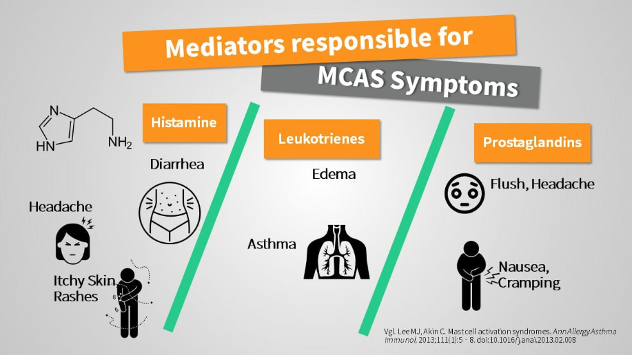
The mediators released by activated mast cells include cytokines like TNF-alpha, interleukin-1, and interleukin-6, which also play a role in inflammatory conditions more broadly. But the main mediators are chemical substances responsible for allergic symptoms, particularly histamine and leukotrienes, along with some pro-inflammatory prostaglandins. [2]
Histamine is probably familiar to anyone dealing with allergies, since antihistamines (anti meaning "against") are medications aimed specifically at counteracting histamine's effects, the ones responsible for allergic symptoms. The histamine released during mast cell activation is also why many classic MCAS symptoms closely resemble those of allergies. Other substances mast cells can release include heparin, a blood-thinning substance, and serotonin. [2]
Serotonin does not just affect mood, it also helps regulate the sleep-wake cycle through its metabolite, melatonin. The general fatigue and brain fog familiar to many people with allergies and mast cell conditions likely has multiple contributing causes, but a connection to serotonin seems plausible, at least in part. [3]
Symptoms of mast cell activation syndrome (MCAS)
Symptoms from "hyperactive" mast cells depend heavily on which mediators are released in each individual patient, which makes them wide ranging and, to some extent, different from person to person. Multiple organ systems are often affected, and which ones depends on the severity and other individual factors. Histamine is often the mediator behind the symptoms that stand out the most.
Symptoms of MCAS include:
- Runny nose, sneezing
- Swelling, for example in the face (angioedema)
- Itching
- Migraine-like headaches
- Severe allergic reactions
- Diarrhea, nausea, and vomiting
- Asthma
- Flushing (reddening of the body)
- Migrating joint pain and inflammation [2]
This list isn not exhaustive, since a single mast cell can release several hundred different mediators, only some of which have been researched and are currently understood. [4] Still, some of the most important and best-understood mediators are histamine, leukotrienes, prostaglandins, tryptase, and heparin, worth taking a closer look at each one.
Histamine-mediated symptoms in MCAS
Histamine is probably the mediator whose effects are the most noticeable and visible. On the skin, it causes an intensely itchy rash known as hives (urticaria). In areas where the skin is very thin, like the eyelids or lips, it often causes swelling.
Edema
This happens because histamine makes blood vessel walls more permeable to fluid, a process doctors call increased vascular permeability. This allows fluid to move from the blood into the surrounding tissue, causing it to swell, a condition known as angioedema (the "angio" part refers to blood vessels, "edema" means fluid buildup in tissue).
Older medical texts often refer to this as Quincke's edema, named after the German physician Heinrich Irenaeus Quincke, who first described it (August 26, 1842 to May 19, 1922).
Migraine
The migraine-like headaches sometimes seen in MCAS are also likely related to increased blood vessel permeability. Within the rigid, bony confines of the skull, the extra fluid entering brain tissue likely creates pressure, which is thought to cause the pain.
Diarrhea
Diarrhea works through the same mechanism, except the fluid leaking from blood vessels enters the intestinal wall and then the gut itself, making stool more watery.
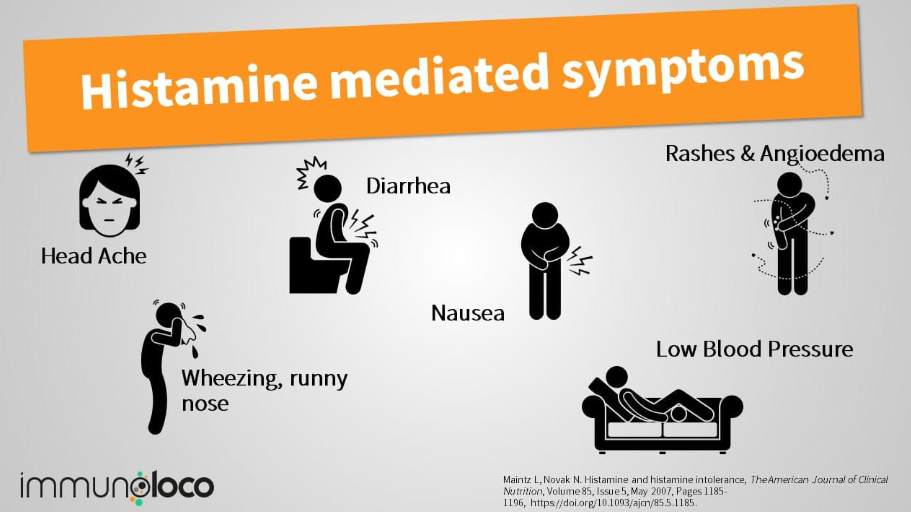
Severe allergic reactions, including airway swelling (again, edema) and circulatory collapse, are also driven by histamine. These are known as anaphylactic reactions, or anaphylaxis. [5]
Anaphylaxis is a life-threatening emergency, marked by difficulty breathing and a drop in blood pressure (caused both by fluid shifting out of the blood vessels and by the vessels widening), often accompanied by intense fear and cold sweats.
Anaphylaxis versus anaphylactoid reactions
While anaphylaxis is a "true" allergic reaction, driven by an excessive immune response to contact with an antigen (the substance someone is allergic to), there are also so-called "pseudoallergies."
These are marked by symptoms that, without lab testing, cannot be distinguished from a true allergic reaction, since many of the same mediators get released. On a biochemical level, though, the actual immune reaction between antigen and antibodies (usually IgE) is missing.
To distinguish the two, these are called anaphylactoid rather than anaphylactic reactions, the suffix "-oid" roughly meaning "similar to."
A classic example of this kind of pseudoallergy is the reaction to non-steroidal anti-inflammatory drugs (NSAIDs) like aspirin, seen in salicylate intolerance.
Prostaglandin-mediated symptoms
Mast cells can also release prostaglandins in significant amounts. Depending on the specific type, their effects vary and can sometimes even be opposite. Some prostaglandins have anti-inflammatory effects, while others promote inflammation.
Prostaglandins form through a complex metabolic pathway that requires specific enzymes called cyclooxygenases. There are two types: COX-1 and COX-2.
Blocking these enzymes, for example by taking non-steroidal anti-inflammatory drugs (NSAIDs) like ibuprofen, aspirin® (acetylsalicylic acid), or diclofenac, reduces the body's prostaglandin production, easing symptoms like headaches and inflammation that these substances mediate.

Prostaglandins are also linked to nausea, abdominal pain, and flushing, the sudden reddening of the body or face. They are also suspected of contributing to the notorious "brain fog". [2]
Leukotriene-mediated symptoms
Alongside histamine, leukotrienes in particular promote mucus production and airway constriction. In sensitive individuals, this can trigger asthma attacks and even bronchospasm, a sudden, cramp-like narrowing of the airways.
Leukotrienes play a major role not just in MCAS but also in conditions like Samter's triad, where patients typically experience more severe asthma attacks, nasal polyps, and aspirin intolerance. [2] [6] For this reason, elevated leukotriene levels are also often seen in MCAS patients who have salicylate (NSAID) intolerance.
The complex effects of these mediators also lead many mast cell patients to experience sinus inflammation (sinusitis) and a runny nose (rhinitis), without any underlying infection. [2] [5]
Other mediators: tryptase, heparin, and cytokines
Tryptase as a marker of mast cell activation
Another substance released by mast cells is the protein tryptase, which is released almost exclusively by mast cells. This happens passively to some extent, continuously and at low levels.
It is also released actively into the bloodstream during activation and degranulation, causing levels to spike sharply before gradually returning to baseline over several hours. This makes tryptase a useful diagnostic marker for estimating the potential severity of allergic reactions. [2] [4]
Heparin as a trigger for bleeding in mast cell conditions
Mast cells can also release heparin, a substance that inhibits blood clotting. Doctors have used it in various forms for years to prevent blood clots, particularly in patients who are largely immobile for extended periods, after surgery, for example.
In MCAS patients, however, the large amounts of heparin released can lead to bleeding in various parts of the body and other complications. On the skin, this shows up as bruising, medically referred to as hematomas. [4]
Serotonin: a contributor to sleep disturbances and brain fog?
Serotonin does not just affect mood, it also helps regulate the sleep-wake cycle through its metabolite, melatonin. The general fatigue and brain fog familiar to many people with allergies and mast cell conditions is likely multifactorial, but a connection to serotonin seems plausible. [3]
This also highlights the close relationship between neurotransmitters and the immune system, and vice versa, a connection that can sometimes be used therapeutically as well.
Interleukin-6, interleukin-1, and TNF-alpha
Mast cells also release cytokines, including interleukin-6, interleukin-1, and TNF-alpha. All of these can promote and trigger inflammation throughout the body. These mediators are especially relevant in rheumatological conditions like rheumatoid arthritis, connective tissue diseases (autoimmune conditions affecting connective tissue), and vasculitis (inflammatory blood vessel disease). MCAS patients sometimes experience migrating joint inflammation or fever as well. [4] [5]
At the same time, these mediators can activate mast cells themselves, which means that once activation begins, it can become self-sustaining rather than resolving on its own. Breaking this cycle is often far from straightforward.
Why MCAS can be difficult to diagnose
The previous symptom list is far from exhaustive, since mast cells release many different mediators that act at many different sites in the body.
Because so many of these symptoms overlap with other conditions, some of which require urgent treatment, doctors and other clinicians often find it difficult to connect the dots between individual symptoms and reach the right diagnosis.
Part of the difficulty also comes from the fact that MCAS diagnosis is not governed by a single, universally agreed standard. Two different diagnostic frameworks currently coexist in the field. The more conservative "consensus-1" criteria (Valent, Akin, Metcalfe, and colleagues) require lab-confirmed evidence of mediator elevation during a flare, most notably a rise in serum tryptase of at least 20% plus 2 ng/mL above baseline, along with a response to mast-cell-targeted treatment. [7] The broader "consensus-2" criteria (Afrin, Molderings, and colleagues) also accept patients whose clinical picture strongly points to mast cell activation even when repeated mediator testing comes back normal, on the basis that tryptase alone may miss many genuine cases. [8]
No international body, including the World Health Organization, has endorsed one framework over the other, and to date no study has directly compared the two approaches head to head. A 2026 six-year follow-up review of the consensus-2 criteria found that their broader approach has improved diagnosis for many patients previously dismissed as having unexplained symptoms or misdiagnosed with a primary psychiatric condition, without the overdiagnosis that some had feared. [9]
References
[1] Kritas SK, Saggini A, Cerulli G, et al. Asthma and Mast Cell Biology. Eur J Inflamm. 2014;12(2):261-265. doi:10.1177/1721727X1401200205
[2] Lee MJ, Akin C. Mast cell activation syndromes. Ann Allergy Asthma Immunol. 2013;111(1):5-8. doi:10.1016/j.anai.2013.02.008
[3] Meeusen R, Watson P, Hasegawa H, Roelands B, Piacentini MF. Central fatigue: the serotonin hypothesis and beyond. Sports Med Auckl NZ. 2006;36(10):881-909. doi:10.2165/00007256-200636100-00006
[4] Afrin LB, Self S, Menk J, Lazarchick J. Characterization of Mast Cell Activation Syndrome. Am J Med Sci. 2017;353(3):207-215. doi:10.1016/j.amjms.2016.12.013
[5] Molderings GJ, Brettner S, Homann J, Afrin LB. Mast cell activation disease: a concise practical guide for diagnostic workup and therapeutic options. J Hematol Oncol. 2011 Mar 22;4:10. doi:10.1186/1756-8722-4-10
[6] Celik G, Bavbek S, Misirligil Z, Melli M. Release of cysteinyl leukotrienes with aspirin stimulation and the effect of prostaglandin E(2) on this release from peripheral blood leucocytes in aspirin-induced asthmatic patients. Clin Exp Allergy J Br Soc Allergy Clin Immunol. 2001;31(10):1615-1622. doi:10.1046/j.1365-2222.2001.01074.x
[7] Valent P, Akin C, Metcalfe DD. Mastocytosis: 2016 updated WHO classification and novel emerging treatment concepts. Blood. 2017;129(11):1420. doi:10.1182/blood-2016-09-731893
[8] Valent P, Akin C, Bonadonna P, et al. Mast cell activation syndrome: Importance of consensus criteria and call for research. J Allergy Clin Immunol. 2018;142(3):1008-1010. doi:10.1016/j.jaci.2018.06.004